Short, clear descriptions of microscope classification and common light microscopy techniques.
 The two main salient features of microscopes are
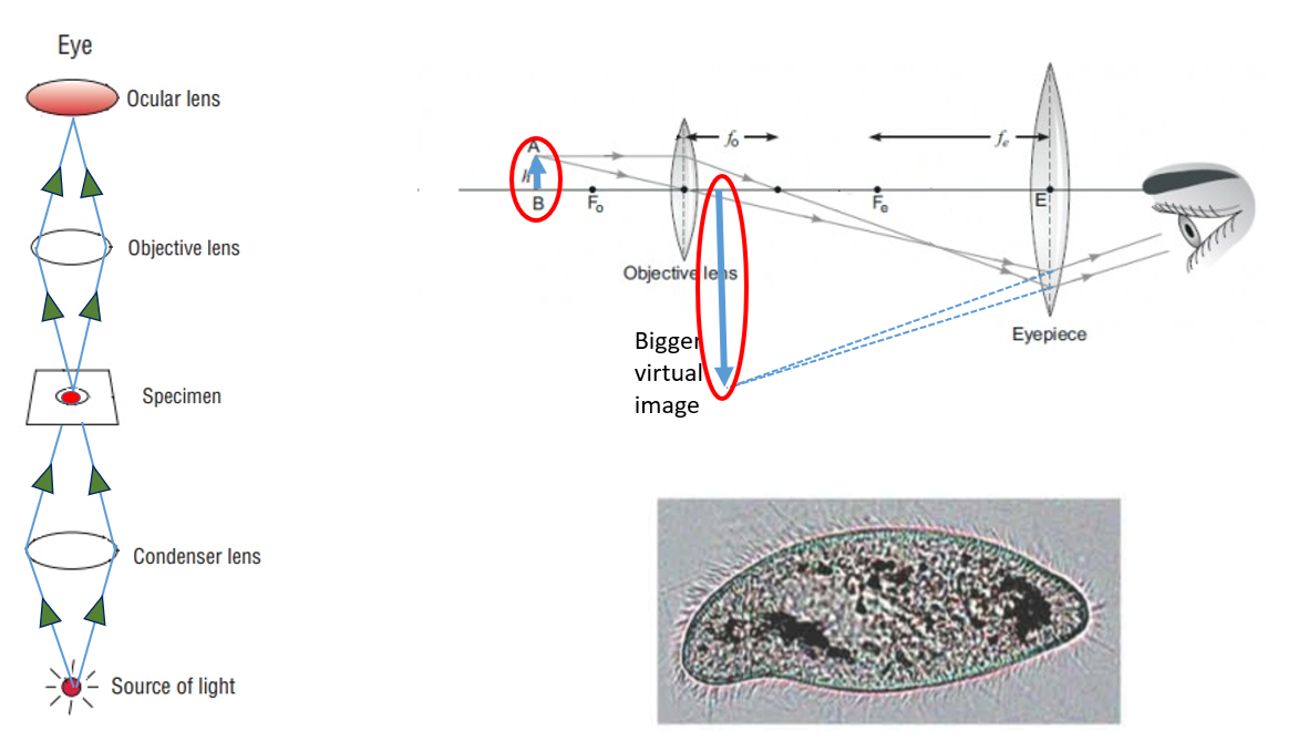
Magnification Power
Magnification Power is the ability to enlarge any object being viewed or it is an ability of the microscope to create a larger view of any object. Magnification power depends on Size and curvature of the lenses.
Total magnification of any microscope is calculate by multiplying the magnification power of objective lens to the ocular lens.
The two main salient features of microscopes are
Magnification Power
Magnification Power is the ability to enlarge any object being viewed or it is an ability of the microscope to create a larger view of any object. Magnification power depends on Size and curvature of the lenses.
Total magnification of any microscope is calculate by multiplying the magnification power of objective lens to the ocular lens.
| Magnification power of Objective lens (A) | Magnification power of ocular lens (B) | A multiplied with B | Total magnification power of microscope |
|---|---|---|---|
| 10 X | 10 X | 100 | 100 times |
| 40 X | 10 X | 400 | 400 times |
| 100 X | 10 X | 1000 | 1000 times |

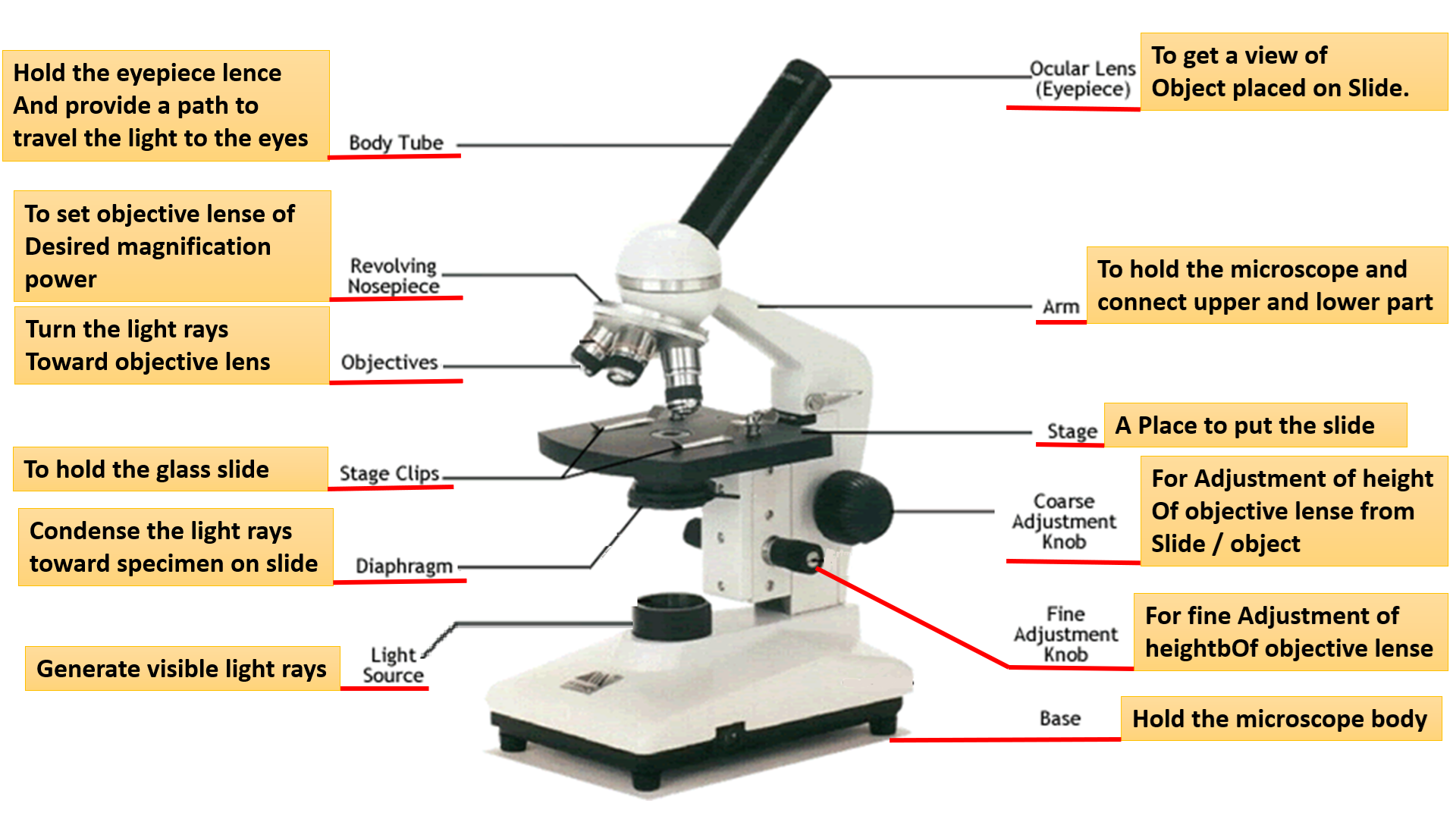
Classification by lens system
Simple Microscope
A simple microscope uses a single lens. There is no combination of two or more lenses.
Compound Microscope
A compound microscope consists of two or more lens elements (for example an objective + an eyepiece) that together magnify the specimen.
Classification by illumination source
Depending on the source of illumination, microscopes can be classified as:
- Light Microscope — uses visible light or ultraviolet light to illuminate the specimen.
- Electron Microscope — uses an electron beam to form the image on a fluorescent or digital display instead of visible light.
| Light Microscopy | Electron Microscopy |
|---|---|
| Bright Field Microscopy | Transmission Electron Microscope (TEM) |
| Dark Field Microscopy | Scanning Electron Microscope (SEM) |
| Phase Contrast Microscopy | — |
Bright Field Microscope
The bright field compound microscope is the most common type used in laboratories. The specimen appears bright and colourful while the background is also bright.
This is a simpler technique because no special lens arrangement is required — most of the light from the source (refracted or unrefracted) reaches the eye, so both image and background appear bright.
Staining is usually required during slide preparation because many microorganisms are colourless and absorb little light. Staining increases the light-absorbing ability of cells and improves contrast and differentiation of cellular structures.
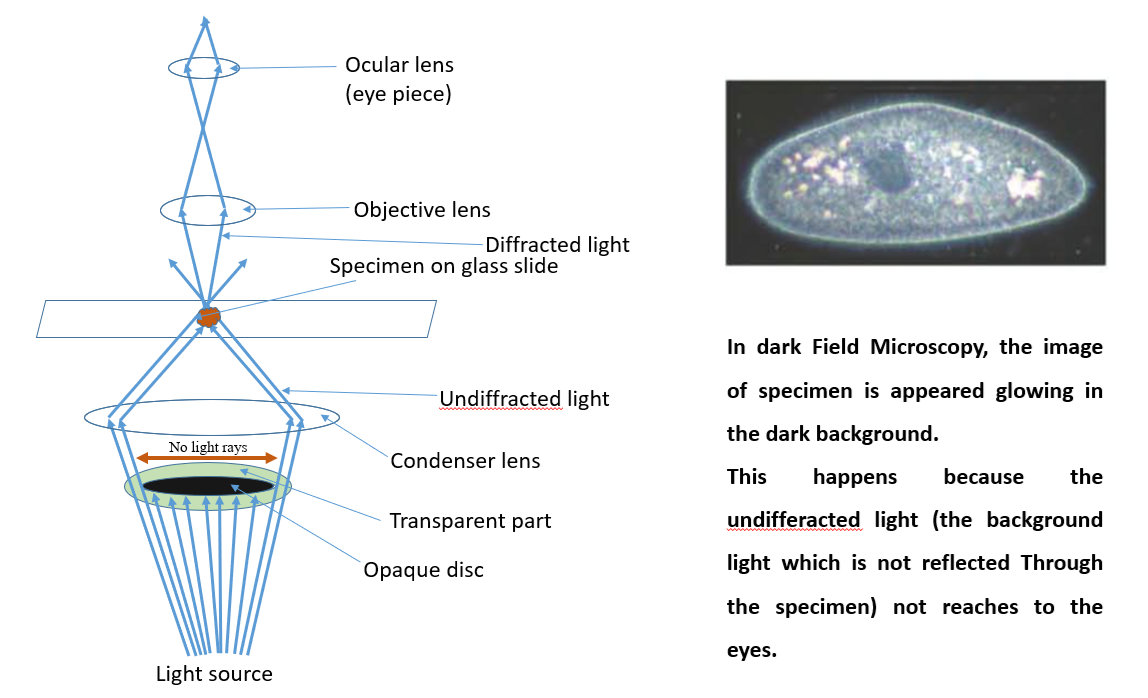
Dark Field Microscopy
In dark field microscopy, only diffracted light (light that interacts with the specimen) is used to form the image; the specimen appears to glow against a dark background.
When the condenser focuses light on the specimen, some rays hit the specimen and are diffracted (their path changes). Others pass undeviated (undiffracted). Undiffracted rays create the dark background, while the diffracted rays produce the specimen image.
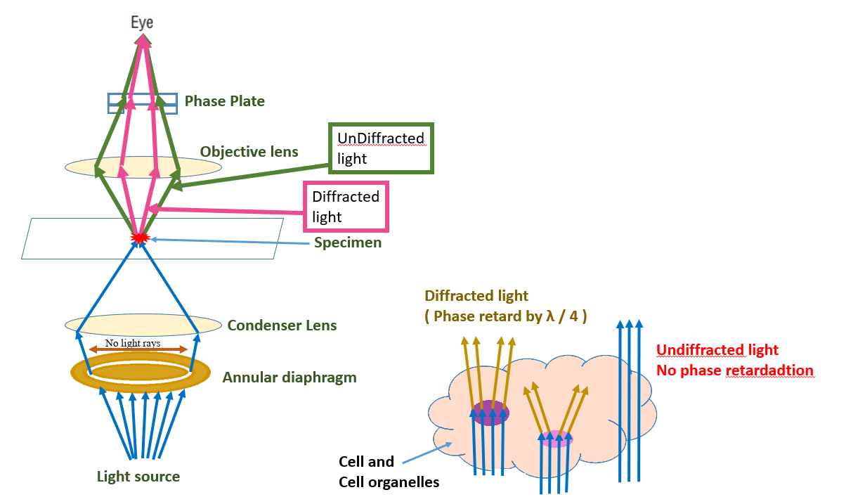
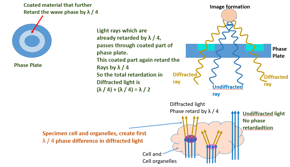
Phase Contrast Microscopy
Phase contrast microscopy differs from bright and dark field techniques. It uses phase differences in light rays to form an image of unstained specimens.
Different cell components have different refractive indices, so as light passes through the cell each component shifts the light's phase. A phase plate above the objective recombines these rays and converts phase differences into amplitude differences, producing a bright image of an unstained specimen against a background that often appears darker or colourful depending on the setup.
Note: in some phase contrast setups a diaphragm blocks part of the light so that undiffracted light does not reach the eye, enhancing contrast.